Pain and performance that never really resolve
You have tried stretches, you have tried strengthening, you have tried yet another new device. Things help a little, then the same limitations creep back. The most common reason this happens is simple and often overlooked: the nervous system and the fascial system are talking past each other. Fascia is richly innervated, capable of transmitting nociceptive and proprioceptive signals, and it remodels in response to load and inflammation. When the conversation between neural circuits and fascial tissues is garbled, the brain builds protective movement patterns that feel “stuck.” Research demonstrates that fascia is densely innervated and can meaningfully influence pain and movement, which means lasting change requires coordinated input to both the tissue and the nervous system.
A chiropractic neurology lens
Chiropractic neurology focuses on assessing functional circuitry—how eyes, vestibular organs, cerebellum, basal ganglia, parietal and frontal cortices, and spinal networks integrate to produce movement and perception. Instead of treating a tight muscle in isolation, we ask: what brain maps and reflex loops are reinforcing this pattern, and what sensory inputs will unlock it? That is the clinical gap MyoSynaptics is designed to fill.
What is MyoSynaptics?
MyoSynaptics is a clinical framework created by Dr. Marc Ellis that combines targeted myofascial assessment and treatment with precisely dosed neurological rehabilitation. In practice, it integrates three pillars:
- Somatotopic mapping and pattern recognition. Practitioners evaluate “neuromyofascial” patterns across kinetic chains—how scars, densification, or adhesions bias joint coupling, ocular and vestibular set points, and cortical body maps.
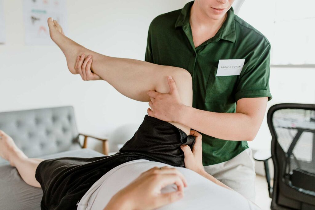
- Tissue-specific myofascial interventions. Hands-on methods are used to alter fascial gliding, fluid dynamics, and mechanoreceptor input. These techniques seek to change signal quality from peripheral receptors rather than “breaking up” tissue.
- Neuro-priming and task integration. Immediately after tissue work, patients perform customized brain-based drills (oculomotor, vestibular, proprioceptive, or cerebellar tasks) to consolidate new maps, improve motor timing, and reduce the likelihood that old protective patterns reassert themselves.
At its core, MyoSynaptics treats fascia as a sensory organ and the brain as the pattern generator that must be re-educated while the tissue input is being updated. This approach is consistent with the scientific understanding that fascia contains abundant proprioceptors and nociceptors and that manual inputs can modulate central processing through mechanotransduction and altered afferent signaling.
How the pieces fit together: From mechanoreceptors to motor maps
When a clinician applies a slow, pressure- and vector-specific fascial input, mechanoreceptors in the tissue fire differently. That change travels through dorsal horn gating, cerebellar and basal ganglia loops, and up to somatosensory and premotor cortices. If, in that exact window, the patient performs a gaze stabilization drill, a head-on-body tracking task, or graded load through a newly liberated range, the nervous system “updates” its model of the limb or segment. Over time, this reduces nociceptive gain, improves joint coupling, and can normalize autonomic arousal that often accompanies persistent pain. Systematic reviews have found that myofascial release can improve pain and certain aspects of function in chronic conditions, while fascial innervation research explains why these inputs are potent. (See references.)
What makes MyoSynaptics different from generic soft-tissue work
- Assessment is neurologically anchored. Practitioners screen oculomotor function, vestibular reflexes, cortical laterality, and cerebellar timing, then correlate those findings with myofascial restrictions.
- Interventions are sequenced to exploit neuroplastic windows. Tissue inputs are followed immediately by drills that target the most plastic node in the circuit (for example, VOR x1 during a newly integrated thoracic rotation pattern).
- Progressions are map-based, not symptom-chasing. The plan advances as sensorimotor discrimination and movement variability improve, not only as pain decreases.
About the founder: Dr. Marc Ellis
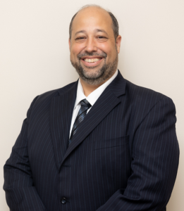 Dr. Marc Ellis is a chiropractic neurologist and soft-tissue specialist who founded MyoSynaptics to codify a neuro-fascial approach to care. He is listed as a founding member of the Fascia Research Society.
His professional background includes early training and certification in neuromuscular therapy, chiropractic education at Life University, and a Master’s degree in Clinical Neuroscience from Parker University (2020). He teaches for the Carrick Institute and has completed extensive postdoctoral study in functional neurology.
Dr. Ellis serves as the clinic director at Georgia Chiropractic Neurology Center, where he and the team integrate myofascial methods with brain-based rehabilitation.
The Fascia Research Society, and how Dr. Ellis is related to it
The Fascia Research Society (FRS) was established to advance the scientific study of fascia and to create an international community where clinicians, educators, and researchers can collaborate. Before the Society’s founding, fascia was often dismissed as “packing material” in anatomy. Over the past two decades, groundbreaking histological, biomechanical, and neurophysiological research has shown that fascia is far from passive. It is richly innervated, it influences proprioception and pain, and it adapts to stress, injury, and movement. The Fascia Research Society was created to bring structure and visibility to this field.
Mission and vision
The Society’s mission is to promote, encourage, and facilitate interdisciplinary research and dialogue on fascia. It provides resources for scientists studying the molecular biology of connective tissue, clinicians investigating manual therapy approaches, and educators teaching anatomy and movement science. The Society envisions fascia as a bridge between disciplines: orthopedics, neurology, sports science, rehabilitation, osteopathy, chiropractic, and beyond.
Dr. Marc Ellis is recognized as a founding member of the Fascia Research Society. This status is significant because it places him among the early clinicians and scientists who saw fascia not as background tissue, but as a vital player in human movement and health. Being part of the Society from its inception shows his commitment to grounding his clinical innovations in research.
This connection also underscores why MyoSynaptics integrates fascia with neurology. The FRS has consistently emphasized the sensory and communicative role of fascia, and Dr. Ellis has taken those principles into clinical practice by pairing fascial inputs with targeted neurological rehabilitation. His involvement in the Society not only keeps him at the forefront of fascia research but also allows him to contribute his own neuro-fascial insights back into the international conversation.
How a MyoSynaptics visit unfolds
- Step 1: Neuro-fascial assessment. Expect eye movement and vestibular screens, positional testing, sensorimotor discrimination tasks, and palpatory mapping of fascial densification along kinetic chains.
- Step 2: Targeted tissue inputs. Slow, specific myofascial vectors improve glide, hydration, and receptor signaling in the precise lines that the assessment uncovered.
- Step 3: Neurological rehabilitation. In combination with the tissue input, your clinician selects specific neurological rehabilitation exercises to consolidate new maps: This can include visual fixation under load, head-on-body tracking, rhythmic stabilization, graded gait exploration and many more.
- Step 4: Reassessment and progression. Changes in gaze metrics, movement variability, range, and load tolerance guide each progression.
When you should consider this approach
- Recurrent pain that returns when you stop doing your exercises.
- Movement limitations that vary with stress, sleep, or visual demand.
- Sensory overload, dizziness, or autonomic symptoms that flare with mechanical strain.
- Post-surgical or scar-related stiffness that does not respond to stretching alone.
- Performance plateaus where strength gains fail to translate into fluid movement.
What success looks like
Success is not just lower pain. It is cleaner joint coupling, smoother eye–head–body coordination, improved movement variability, steadier autonomic tone, and the confidence that you can load tissue without tripping an alarm. Patients often report that everyday tasks feel less effortful and that training feels “available” again because the brain’s prediction of movement cost has changed.
Why this matters now
Fascia research has matured considerably. Histological and immunohistochemical work shows dense innervation of deep fascial layers, including nociceptors that can upregulate in inflammatory states. Systematic reviews and meta-analyses suggest that manual approaches aimed at fascia can improve pain and function in several conditions, though effect sizes vary and study quality is mixed. The practical takeaway is clear: when manual inputs are paired with precise, brain-based rehabilitation, the central nervous system is more likely to adopt new, efficient patterns.
If you or someone you love is experiencing symptoms that you are not finding any explanation for or relief from and you would like to learn how chiropractic neurology and MyoSynaptics can help, contact the team at Georgia Chiropractic Neurology Center today. We look forward to hearing from you.
Written by Sophie Hose, DC, MS, DACNB, CCSP
Peer-Reviewed References
- Van der Kolk, B. A. (2014). The body keeps the score: Brain, mind, and body in the healing of trauma. Viking.
- Lopez, C., Blanke, O., & Mast, F. W. (2012). The human vestibular cortex revealed by coordinate-based activation likelihood estimation meta-analysis. Neuroscience, 212, 159–179.
- Smith, P. F., & Darlington, C. L. (2013). Personality changes in patients with vestibular dysfunction. Frontiers in Human Neuroscience, 7, 678.
- Porges, S. W. (2011). The polyvagal theory: Neurophysiological foundations of emotions, attachment, communication, and self-regulation. W.W. Norton & Company.
- Yardley, L., Beech, S., Zander, L., Evans, T., & Weinman, J. (1998). A randomized controlled trial of exercise therapy for dizziness and vertigo in primary care. British Journal of General Practice, 48(429), 1136–1140.