
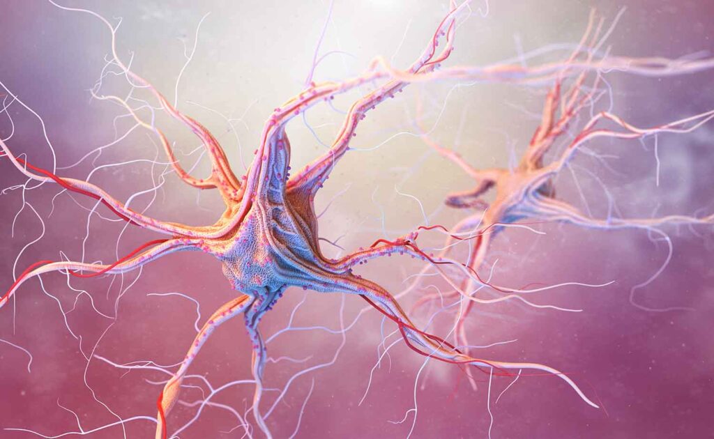
Imagine if your nervous system suddenly began expressing movement patterns that should only be seen in infancy.
A gentle stroke along the foot causes the toes to fan upward.
A light touch to the palm causes an involuntary grasp.
A tap on the face triggers an abnormal response.
To most people, these reflexes seem unrelated to daily life. To neurologists, however, they can provide valuable information about how the brain and nervous system are functioning.
These responses are known as pathological reflexes.
Despite the name, the reflexes themselves are not necessarily the problem. Rather, they are often signs that something has changed in the nervous system’s ability to regulate and inhibit lower-level neurological activity.
For chiropractic neurologists, pathological reflexes can serve as important clues during a neurological examination, helping identify areas of dysfunction and guiding individualized rehabilitation strategies.
Most importantly, these reflexes remind us that healthy neurological function depends not only on activating the right pathways, but also on inhibiting the wrong ones.
Searching for Answers About Neurological Function
Many individuals seek neurological care because they notice changes that are difficult to explain.
They may experience:
- Balance problems
- Coordination difficulties
- Weakness
- Changes in walking patterns
- Reduced dexterity
- Neurological symptoms after a concussion
- Persistent dizziness
- Cognitive changes
- Post-stroke deficits
- Progressive neurological symptoms
Sometimes these symptoms develop suddenly.
Other times they emerge gradually.
In many cases, neurological examination provides important clues that imaging alone cannot reveal.
One of the most valuable tools available to clinicians is the reflex examination.
Reflexes allow healthcare providers to evaluate how different levels of the nervous system communicate with one another.
When certain reflexes appear unexpectedly, they may signal that normal neurological inhibition has been disrupted.
The Nervous System Depends on Inhibition
Most people think of the nervous system in terms of activation.
Muscles contract.
Nerves fire.
The brain sends signals.
While activation is important, inhibition is equally critical.
In fact, much of healthy brain function depends on the ability to suppress inappropriate responses.
The brain constantly filters information.
It decides which signals should be amplified and which should be suppressed.
Without this process, movement would become inefficient and behavior would become chaotic.
Pathological reflexes provide a fascinating example of this principle.
Many of these reflexes are present early in life as part of normal neurological development.
As the brain matures, higher cortical centers gradually gain control over lower reflex pathways.
The reflexes do not disappear completely.
Instead, they become inhibited.
This distinction is important.
The circuitry remains present.
The brain simply keeps it under control.
When higher levels of the nervous system lose their inhibitory influence, these reflexes may become visible again.
What Are Pathological Reflexes?
Pathological reflexes are involuntary responses that are generally absent in healthy older children and adults.
Their presence often suggests dysfunction involving the central nervous system, particularly the corticospinal tract or other upper motor neuron pathways.
These reflexes are considered “released” because the normal inhibitory influence from higher brain centers has been reduced.
Rather than representing new neurological activity, they often represent old neurological programs becoming unmasked.
The reflex was always there.
The brain was simply suppressing it.
When that suppression weakens, the reflex becomes observable again.
Common Pathological Reflexes
Babinski Sign
Perhaps the most well-known pathological reflex is the Babinski sign.
When the sole of the foot is stroked, the big toe extends upward while the other toes fan outward.
In infants, this response is normal.
In adults, it often suggests dysfunction involving upper motor neuron pathways.
The Babinski sign may be observed in conditions affecting the corticospinal tract, including:
- Stroke
- Multiple sclerosis
- Brain injury
- Spinal cord injury
- Neurodegenerative disease
Hoffman Reflex
The Hoffman reflex is often considered the upper-extremity equivalent of the Babinski sign.
A flick of the middle finger produces involuntary flexion of the thumb or index finger.
A positive Hoffman response may indicate increased excitability within corticospinal pathways.
It can sometimes be seen in:
- Cervical spinal cord disorders
- Multiple sclerosis
- Upper motor neuron lesions
- Certain neurological disorders
Grasp Reflex
When the palm is stimulated, the hand involuntarily closes around the object.
This reflex is normal in infancy but typically disappears as the frontal lobes mature.
Its reappearance in adults may indicate frontal lobe dysfunction.
Snout Reflex
Light stimulation around the mouth causes puckering or protrusion of the lips.
This reflex may be associated with frontal lobe disinhibition.
Palmomental Reflex
Stimulation of the palm causes contraction of muscles around the chin.
While occasionally present in healthy individuals, exaggerated responses may indicate neurological dysfunction.
What Pathological Reflexes Tell Us
A pathological reflex does not provide a diagnosis by itself.
Instead, it acts as a clue.
Neurologists often describe these reflexes as signs of “release.”
Higher brain centers normally exert inhibitory control over lower neurological circuits.
When this control becomes impaired, primitive or pathological reflexes may emerge.
Think of it like a vehicle’s braking system.
The engine may not have changed.
The accelerator may not have changed.
But if the brakes become less effective, behaviors that were previously controlled begin to emerge.
Similarly, pathological reflexes often indicate that inhibitory systems are no longer functioning optimally.
This can occur because of injury, disease, degeneration, inflammation, or altered neurological function.
Conditions in Which Pathological Reflexes May Appear
Pathological reflexes can emerge in a variety of neurological conditions.
These may include:
Stroke
Damage to cortical motor pathways frequently results in loss of inhibitory control.
This often leads to the appearance of upper motor neuron signs, including pathological reflexes.
Traumatic Brain Injury
Moderate and severe brain injuries can disrupt frontal lobe and corticospinal function, allowing previously inhibited reflexes to reappear.
Multiple Sclerosis
Demyelination within the central nervous system can impair communication between higher and lower neurological centers.
Parkinsonian Syndromes
Certain neurodegenerative conditions may involve the emergence of frontal release signs.
Dementia
Some forms of dementia, particularly those affecting frontal lobe networks, may be associated with primitive and pathological reflexes.
Spinal Cord Disorders
Damage to descending motor pathways may result in pathological reflex expression below the level of injury.
Cerebral Palsy
Depending on the pattern and severity of neurological involvement, retained primitive and pathological reflexes may be present.
Neurodegenerative Diseases
Conditions such as amyotrophic lateral sclerosis (ALS) and other upper motor neuron disorders may demonstrate pathological reflexes as part of the clinical picture.
A Chiropractic Neurology Perspective
Chiropractic neurology focuses on evaluating functional activity within the nervous system.
Pathological reflexes are valuable because they provide objective information about how different regions of the brain and nervous system are communicating.
Rather than viewing a reflex as an isolated finding, chiropractic neurologists evaluate it within the broader context of: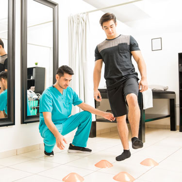
- Balance
- Eye movements
- Coordination
- Gait
- Posture
- Cognitive function
- Sensory processing
- Motor control
A pathological reflex may help identify which neurological networks require further investigation.
For example, the presence of frontal release signs may prompt additional assessment of frontal lobe function.
An abnormal Babinski response may encourage closer examination of corticospinal pathways.
These findings help create a more complete picture of neurological health.
Supporting Neurological Inhibition Through Neuroplasticity
One of the most important concepts in chiropractic neurology is neuroplasticity.
Neuroplasticity refers to the nervous system’s ability to adapt, reorganize, and strengthen neural connections.
Because pathological reflexes often reflect impaired inhibition, rehabilitation strategies may focus on improving the function of the networks responsible for that inhibition.
Depending on the individual’s condition, chiropractic neurological rehabilitation may include:
- Balance training
- Vestibular rehabilitation
- Eye movement therapies
- Coordination exercises
- Gait training
- Sensory integration exercises
- Cognitive rehabilitation
- Dual-task training
- Postural exercises
- Motor control training
The goal is not simply suppressing a reflex.
The goal is improving the efficiency of the neurological systems responsible for regulating that reflex.
As higher brain centers function more effectively, inhibitory control may improve.
This reflects one of the central principles of neurological rehabilitation:
Improving function at higher levels of the nervous system can influence behavior at lower levels.
Why These Reflexes Matter
Pathological reflexes are important because they provide objective evidence of neurological function.
Unlike symptoms, which are subjective, reflexes can often be observed and measured directly.
They help clinicians identify:
- Changes in neurological status
- Areas of dysfunction
- Progression of disease
- Response to rehabilitation
- Integrity of motor pathways
They also remind us that neurological health involves balance.
The brain must know when to activate and when to inhibit.
Both are essential.
Understanding What the Nervous System Is Trying to Tell Us
Pathological reflexes are not simply unusual examination findings.
They are messages.
They tell us something about how the nervous system is functioning.
More specifically, they often reveal that the balance between activation and inhibition has changed.
The reflex itself is rarely the primary problem.
Instead, it is evidence that higher neurological systems may not be exerting their usual level of control.
For chiropractic neurologists, these findings provide valuable information that helps guide assessment and rehabilitation.
By identifying where communication has become disrupted, clinicians can develop strategies aimed at improving neurological function through targeted stimulation and neuroplasticity-based rehabilitation.
Ultimately, pathological reflexes remind us of an important neurological truth:
Healthy brain function is not just about turning systems on.
It is also about knowing when to keep them turned off.
If you or someone you love is experiencing unexplained neurological symptoms and you would like to learn how chiropractic neurology can help, contact the team at Georgia Chiropractic Neurology Center today. We look forward to hearing from you.
Written by Sophie Hose, DC, MS, DACNB, CCSP
Peer-Reviewed References
- Joseph Babinski. Sur le réflexe cutané plantaire dans certaines affections organiques du système nerveux central. Comptes Rendus de la Société de Biologie. 1896.
- Michael Aminoff. Clinical Neurophysiology and neurological examination of pathological reflexes.
- Adams and Victor’s Principles of Neurology. Chapters on upper motor neuron syndromes and pathological reflexes.
- Roger P. Simon et al. Clinical Neurology. Evaluation of primitive and pathological reflexes.
- Richard S. Snell. Clinical Neuroanatomy. Discussion of corticospinal inhibition and reflex control.
- John C. Eccles. Studies on inhibition, motor control, and descending neurological pathways.