Written by Sophie Hose, DC, MS, CCSP
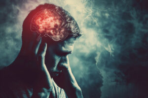
Concussions, or mild traumatic brain injuries (mTBI), are injuries that involve an impact to the head or body. Concussions are typically associated with a change in brain function. They are the most common form of traumatic brain injuries, accounting for about 86% of all TBI cases. Falls make up the most common form of concussions in the general population, followed closely by motor vehicle crashes. Growing concerns are also rising concerning sport-related concussions, with the CDC reporting between 1.6 and 3.8 million cases each year in the US alone.
 Statistics explain the increasingly common demand for improved concussion prevention, education, recognition, and treatment, mTBIs get underreported and not treated appropriately. However, new research suggests long-term, lasting effects even after a single injury.
Our backgrounds, including cultural norms, education, life experiences, social environments, and many more, make our brains unique. With no two brains having the same structure and physiology, no two brain injuries are the same. Coming to this realization makes concussion care more challenging. As symptoms, time spans for recovery, and successful treatment approaches vary from person to person.
Standardized treatment protocols do not meet the injured person’s exact needs. In many cases, signs and symptoms can be missed in regular concussion screenings. The most common test is the SCAT-6 testing battery. If a patient does not recover within the 7-10 days considered ‚normal recovery time, healthcare providers are often at a loss regarding what else to do.
Overall, there are five different types of concussions:
- Cognitive
- Vestibular
- Oculomotor
- Headache/migraine
- Anxiety/mood
These subtypes are fluid and can shift between each other. Concussions cannot be classified according to just one subtype, as patients present with clusters of different symptoms from different subtypes. It is of note, however, that the two most common types are concussions affecting cognition and oculomotor function (eye function).
March is National Brain Injury Awareness Month. We will be highlighting different possible consequences of concussions as related to multiple areas of the brain..
The Frontal Lobe
The frontal lobes are the largest lobes of the human brain and are located directly behind the forehead, a common injury site for head traumas. This makes them the most commonly affected region of injury in traumatic brain injury, according to the Acquired Brain Injury Outreach Service of the Queensland Government in Australia. The frontal lobes is highly involved in executive function and motor output.
The executive function entails various processes that are easily grouped as the skills that enable us to live in a society: Decision making, planning, learning, initiation and control of responses to outside stimuli, and many more. In short, the frontal lobes control our behavior and our emotions. The frontal lobes contain the motor strip, an area of the brain where movements are planned and their execution is initiated. Because of this broad range of functions, an injury to the frontal lobes can cause various symptoms. Common symptoms following a concussion affecting the frontal lobes include:
- changes in personality
- inability to concentrate
- changes in memory
- depression
- anxiety
- trouble moving certain body parts
- difficulty planning movements
- pain syndromes (including headaches)
- light sensitivity
- fatigue
- lack of motivation
- irritability
- restlessness
- drowsiness
- trouble speaking
- mood changes
- difficulty keeping track of tasks
Given the wide variety of symptoms associated with frontal lobe concussions, these kinds of mTBIs can be classified as any one of the five subtypes of concussions mentioned above, depending on the unique combination of symptoms.
Concussions are treated as functional deficits
Within the field of chiropractic neurology, concussions are regarded and treated mainly as functional deficits. In most cases, little to no actual structural damage was sustained from the original injury (such as brain bleeds, open wounds or extreme swelling) and the symptoms that follow the concussion are suspected to be due to other reasons.
These include:
- local short-term swelling of the injured brain tissue (edema)
- changes in blood flow to the area (and therefore changes in the delivery of oxygen and nutrients, as well as decreased removal of waste products and inflammatory markers), changes in the integrity of the blood-brain barrier
- local inflammation
- changes in the internal cell transportation mechanisms of the neurons and
- changes in the biochemical makeup of the neurons, to name a few.
Researchers continue to this day to explore the potential mechanisms of the pathology of concussions, as the complexity and variety of these kinds of injuries make it challenging to come up with a single congruent model.
A recent study by Lemme et al. explored the association between post-traumatic headaches following a concussion and the persistence of concussion symptoms. The researchers used two groups of young patients who had sustained an mTBI, one group whose headache symptoms had resolved, and one group whose symptoms were persistent. They compared self-reported pain scores and MRI and functional MRI data of cortical thickness and subcortical volume of the whole brain.
The results showed that a core network of brain regions (including the frontal lobes) that are involved in the pain response of the patients is altered in individuals who experienced post-traumatic headaches, whether they are resolved or persistent.This means that even though the patient might not be actively experiencing the pain, their brain function is still altered and might cause further symptoms. The researchers, therefore, concluded that people suffering from post-traumatic headaches may experience ongoing functional abnormalities, which might lead to symptom chronification.
This is congruent with research performed by the National Institute on Aging in collaboration with other institutes. Their researchers found that older individuals with a history of one or more concussions had more significant brain atrophy (shrinking of brain tissue) of the frontal and temporal lobe regions. They concluded that concussions can produce long-lasting structural and functional changes in the brain and that many of the reported short-term effects of concussions may still be apparent later in life.
There is more and more discussion surfacing about the potential long-term effects of concussions, especially as the knowledge about chronic traumatic encephalopathy (CTE) grows and more and more professional athletes make the news in regards to diagnosed concussions or even ending their careers early due to head injuries.
Impact of Concussions on Motor Learning in Athletes: Insights from Johns Hopkins Study
A study performed on athletes from Johns Hopkins University who participate in both contact and non-contact sports found that abnormal motor learning (learning new skills that are either sport-related or skills in everyday life), and especially retention of new skills, was associated with abnormal neurophysiological changes in the motor area of the frontal lobes. These findings were present as early as two weeks following the initial injury and persisted years after the last concussion. Furthermore, older athletes might have an even higher chance of developing abnormal motor learning following a concussion later in life.
Research has also shown that concussions involving the frontal lobe can lead to longer reaction times, less hand-eye coordination, poor decision-making, slower visual processing speeds, reduced stress regulation, and difficulty concentrating and sustaining attention. These persistent neuropsychological deficits can be challenging to properly assess, as they may be more subtle than what can be observed with available computerized testing batteries (like, for example, the SCAT 6, the SWAY test, the ImPACT test, and others).
This can be especially challenging for athletes as it not only makes their return to play more difficult but also increases their chance of suffering from repeated concussions should they return before their deficits are rehabilitated to a sufficient extent.
In conclusion, there have been a lot of changes in the perception of and education around concussions in the past few years. More and more research is emerging, which helps healthcare providers worldwide to increase their knowledge base to improve concussion prevention, detection, and treatment. A lot of work remains to be done to protect not only athletes but anyone from the potentially life-changing effects of a mild traumatic brain injury.
Take Control of Your Brain Health with Georgia Chiropractic Neurology Center
As chiropractic neurologists, we strive to assess the integrity of each patient’s brain function in as much detail as possible. This lets us create a treatment plan tailored to their specific situation. At Georgia Chiropractic Neurology Center, each patient undergoes extensive assessments of their autonomic nervous system, oculomotor system (eye function), balance, cognition, and a battery of neurological tests highlighting different brain areas’ functions. Combined with a thorough history and multiple questionnaires. Asking the patient for their perception of their symptoms gives us a detailed overview of the state of their nervous system. From there, the team works together to ensure that the patient can recover as well as possible while continuously observing for any changes in their nervous system.
If you or someone you love has suffered a concussion and would like to learn how chiropractic neurology can help, contact the team at Georgia Chiropractic Neurology Center today. We look forward to hearing from you.
Sources:
-CDC
-Mayo Clinic
-Queensland Health
-Cantarero G, Choynowski J, St Pierre M, Anaya M, Statton M, Stokes W, Capaldi V, Chib V, Celnik P. Repeated Concussions Impair Behavioral and Neurophysiological Changes in the Motor Learning System. Neurorehabil Neural Repair. 2020 Sep;34(9):804-813. doi: 10.1177/1545968320943578. Epub 2020 Jul 29. PMID: 32723160; PMCID: PMC7501144.
-June D, Williams OA, Huang CW, An Y, Landman BA, Davatzikos C, Bilgel M, Resnick SM, Beason-Held LL. Lasting consequences of concussion on the aging brain: Findings from the Baltimore Longitudinal Study of Aging. Neuroimage. 2020 Nov 1;221:117182. doi: 10.1016/j.neuroimage.2020.117182. Epub 2020 Jul 20. PMID: 32702483; PMCID: PMC7848820.
-Poltavski D, Bernhardt K, Mark C, Biberdorf D. Frontal theta-gamma ratio is a sensitive index of concussion history in athletes on tasks of visuo-motor control. Sci Rep. 2019 Nov 26;9(1):17565. doi: 10.1038/s41598-019-54054-9. PMID: 31772237; PMCID: PMC6879532.
-Lemme J, Holmes S, Sibai D, Mari J, Simons LE, Burstein R, Zurakowski D, Lebel A, O’Brien M, Upadhyay J, Borsook D. Altered Brain Network Connectivity Underlies Persistent Post-Traumatic Headache following Mild Traumatic Brain Injury in Youth. J Neurotrauma. 2021 Jun 15;38(12):1632-1641. doi: 10.1089/neu.2020.7189. Epub 2021 Jan 8. Erratum in: J Neurotrauma. 2021 Sep 1;38(17):2486. PMID: 33183144; PMCID: PMC8165471.